Optogenetics gives neuroscientists something electrodes never could: the ability to switch a genetically chosen population of neurons on or off with a pulse of light. The technique works by introducing a gene for a light-sensitive protein—an opsin—into specific neurons, so that those cells, and only those cells, respond to illumination. Shine the right wavelength and the targeted neurons fire; shine another and they fall silent. As a review of the field's molecular tools puts it, progress in understanding brain circuits has historically depended on new technologies that advance the ability to observe and control neuronal activity, allowing researchers to dissect the workings of circuits underlying natural behavior and to see how they become dysfunctional in disease.
The system has three core stages. First, neurons are genetically targeted to express an opsin. Second, light is delivered to the brain region where those neurons sit. Third, the opsin converts that light into a change in the cell's electrical state—either excitation or inhibition—depending on which protein was chosen. The rest of this article walks through those stages and the two workhorse proteins that make bidirectional control possible.
Stage 2: Delivering Light to the Target Region
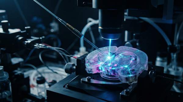
Once a neuron manufactures an opsin and embeds it in its membrane, the cell is primed but inert. Nothing happens until light of the opsin's preferred wavelength reaches it. In a living brain, that means the researcher must route illumination to the target region—typically through an optical fiber implanted above the area of interest, or via a light source positioned over cells in a slice or culture preparation. The physics of the delivery hardware is a discipline of its own; what matters mechanistically is that the light must match the opsin's activation spectrum and reach the expressing cells at a usable intensity.
Wavelength is not a detail—it is the address label of the experiment. Each opsin responds to a particular band of the visible spectrum, and researchers exploit these differences deliberately. Because different opsins are driven by different colors, light itself becomes a selective instrument: one population of cells can be activated while a second population in the same tissue, carrying a different opsin, stays untouched. The choice of wavelength therefore completes the specificity that genetics began, narrowing control from a brain region down to the cells that were actually engineered to respond.
Stage 3: Bidirectional Control of Neural Activity
When light strikes the opsin, the protein changes shape and either opens an ion channel or drives an ion pump. Ions then move across the membrane, and the cell's electrical potential shifts. Which direction it shifts—and therefore whether the neuron is activated or silenced—depends entirely on which opsin was installed. This is the payoff of the whole pipeline: a causal, reversible lever on neural activity.
The two directions of control are complementary in a way that mirrors how circuits are actually studied. Activating a population tests whether those neurons are sufficient to produce a behavior or a signal; silencing them tests whether they are necessary. According to a review of light-driven silencing tools, blue-light driven ChR2 can be co-expressed with yellow- or red-light driven silencers like Halo within a single cell, allowing for both depolarization and hyperpolarization of the targeted neuron using blue and yellow light, respectively. In other words, a single engineered neuron can be pushed in both directions simply by changing the color of the light.
Excitatory Control: Channelrhodopsin-2 (ChR2)
Channelrhodopsin-2 is the standard tool for turning neurons on. According to Emily Ferenczi and colleagues, writing in Frontiers in Physiology in 2019, ChR2 is a non-selective cation channel, permeable to protons far more than sodium ions, then potassium ions, and only weakly to calcium ions under physiological conditions. When the channel pore opens through a sequence of molecular changes initiated by photon absorption, the resulting net inward flow of positive ions produces a depolarizing photocurrent. If that depolarization pushes the excitable membrane past its firing threshold, the neuron generates an action potential.
Wild-type channelrhodopsin has a peak activation spectrum of 470 nanometers—blue light—but the toolkit has since expanded. The same review notes that naturally occurring and engineered variants now cover a wide range of the visible spectrum, from more blue-shifted to red-shifted versions. One early red-shifted variant, VChR1, is maximally activated by green light of roughly 530 nanometers but retains strong photocurrents in amber light at 590 nanometers—a wavelength that produced no measurable depolarization in ChR2-expressing cells. That spectral separation is what lets researchers drive two different opsin populations independently within the same preparation.
Inhibitory Control: Halorhodopsin (NpHR)
Halorhodopsin, also called NpHR, is the mirror-image tool: a light-activated chloride pump used to quiet neurons. Rather than opening a passive channel, NpHR actively transports chloride ions into the cell when illuminated. The influx of negatively charged ions increases the intracellular concentration of negative charge, hyperpolarizing the neuronal membrane and pushing it away from its firing threshold. The result is inhibition—the targeted neuron is effectively silenced for as long as the light is on.
NpHR's activation spectrum sits at the other end of the visible range from ChR2's. It is driven by yellow light, specifically at 589 nanometers, and can also be driven by red light. That separation is what makes the combined experiments described above possible: a neuron expressing both proteins can be depolarized with blue light and hyperpolarized with yellow, giving the experimenter a two-way switch over a single identified cell. The same color logic extends across silencing tools more broadly—one silencing protein is quieted by blue light but not red, while Halo-expressing neurons are quieted by red light and not blue—so the choice of opsin and wavelength together define exactly which cells respond.
Optogenetic System Workflow: From Gene to Neural Control
The table below traces the complete causal sequence of an optogenetic experiment, from the initial genetic modification to the final change in firing. Follow any row left to right to see how each stage sets up the next.
| Stage | Key Component | Mechanism |
|---|---|---|
| 1. Genetic targeting | Opsin gene delivered to chosen neurons | Only the targeted cells come to express the light-sensitive protein in their membranes |
| 2. Light delivery | Wavelength matched to the opsin | Blue light (~470 nm) reaches ChR2-expressing cells; yellow light (589 nm) or red light reaches NpHR-expressing cells |
| 3a. Excitatory outcome | ChR2 cation channel | Photon absorption opens the pore; net inward flow of positive ions depolarizes the membrane and triggers action potentials |
| 3b. Inhibitory outcome | NpHR chloride pump | Light drives chloride ions into the cell, hyperpolarizing the membrane and silencing the neuron |
Read as a chain, the model shows why specificity compounds at each step: genetics restricts which cells carry the protein, wavelength restricts which of those cells respond at a given moment, and the opsin's ion mechanism determines the direction of the effect. Break any link—no gene expression, no light, no matching wavelength—and the control fails.
Applying Optogenetics: Precision in Neural Circuit Dissection
Researchers can precisely dissect neural circuit function by selectively activating or inhibiting specific neuron populations using optogenetics. The observable signal is the specific change in neuronal firing patterns—depolarization or hyperpolarization—in response to targeted light delivery, indicating successful manipulation of circuit activity. That readout is the test of every design choice described here: if blue light over a ChR2-expressing population produces the expected burst of action potentials, and yellow light over an NpHR-expressing population produces the expected silence, the causal role of those cells in the circuit under study can be established directly rather than inferred.