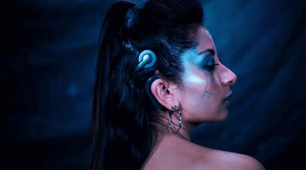
Sensory neuroprosthetics all work from the same basic premise: when the natural pathway for a sense is damaged, an implanted device can deliver electrical stimulation to a downstream part of the nervous system and evoke a usable percept. But where each device plugs into that system — and what kind of sensation it produces — varies enormously. A cochlear implant reaches the auditory nerve, a retinal implant targets surviving cells inside the eye, and a somatosensory prosthetic stimulates the peripheral nervous system or the brain itself. Understanding those interface differences explains why the three technologies deliver such different results: speech understanding for cochlear recipients, deliberately limited visual percepts for retinal implant users, and paresthetic touch feedback for people with prosthetic limbs.
Introduction to Sensory Neuroprosthetics
The three device classes compared here occupy very different stages of maturity. Cochlear implantation is a well-established clinical treatment, supported by policy documents that describe consistent effectiveness in speech reception and sound localization. Retinal implants are a more experimental field, with device designs split between two competing anatomical placements. Somatosensory prosthetics are the newest of the three, emerging from research that has largely demonstrated feasibility rather than everyday clinical use.
What unites them is the strategy of substituting electrical stimulation for a lost biological transduction step. Each device chooses a different target along its sensory pathway, and that choice determines what the patient perceives. The closer the stimulation lands to the brain's own processing hierarchy, the more the resulting percept depends on higher-level neural interpretation — and the more the outcome varies from person to person. This article compares the three technologies on exactly those dimensions and keeps to the general principles described in the cited research rather than any specific commercial device.